Brain scans
Brain scans can be useful in diagnosing epilepsy. They are also used to work out if a person may be suitable for surgery,when it is necessary to confirm that seizures are arising from one part of the brain and that it is safe to remove this part. This requires many tests including MRI brain scans.
A brain scan may help to find the cause of your seizures. The two common types of brain scan are Magnetic Resonance Imaging (MRI) and Computerised Axial Tomography (CT or CAT).
The scan produces pictures of the brain which might show a physical cause for epilepsy, such as a scar on the brain. But for many people a brain scan does not show a cause for their seizures, and even if no physical cause is seen, the person may still have epilepsy.
Magnetic Resonance Imaging (MRI) scan
An MRI scan looks at the structure of the brain and may help to find the cause of your epilepsy. During the scan, detailed pictures are produced using strong magnetic fields. Because of the magnetic fields, metal objects in or near the machine can affect, or be affected by, the machine.
Before having an MRI scan you will need to remove any metal objects such as jewellery, hearing aids, coins or keys. If you have a heart pacemaker or any surgical implant that contains metal you may not be able to have an MRI scan.
The scanner makes a loud knocking noise, so before it starts you will be given earplugs to wear. You will also be given a buzzer to hold so you can let the technician know if you are uncomfortable or feeling unwell during the scan.
The technician is usually on the other side of a window in another room during the scan. There is an intercom so you can talk to them and a mirror so you can see them. You may be able to have someone in the room with you during the scan.
Having an MRI scan to help diagnose epilepsy usually takes about 30 minutes. During the scan you will lie on a platform which slides into the scanner (a bit like going into a tunnel).
It is important to lie still during the scan so that the machine can take clear pictures of your brain. An MRI scan is usually a series of short scans with breaks in between, rather than one long scan. Between each scan the technician might use the intercom to check that you are ok.
Computerised axial tomography (CT or CAT scan)
Some people may have a CT scan if they are not able to have an MRI scan. This might be because they have a heart pacemaker, if they might need to have an anaesthetic to have an MRI or if information about what might be causing their seizures is needed quickly.
CT scans use X-rays to take images of the brain. (CT scans are not suitable if you are pregnant because the X-rays could affect an unborn baby).
Images from a CT scan are less detailed than those from MRI scans. During a CT scan you lie on a couch which slides into the scanner. Unlike MRI scanners, CT scanners do not make a loud noise.
New MRI techniques
Despite advances in technology, around 30 per cent of people with seizures arising from one part of the brain have completely normal MRI scans. Further investigation with other types of scan is expensive, may involve radiation exposure and is not available in most places, unlike MRI. If imaging cannot identify the source of seizures, recording wires may be placed directly into the brain and on to the brain surface. This is available in only a few places, is invasive, very expensive and carries some risk.
We are assessing several new MRI techniques to determine which are helpful in identifying the source of seizures in people with severe epilepsy and normal standard scans. We work with the UCL Centre for Medical Image Computing to develop new ways to analyse the scans by computer to detect subtle abnormalities not seen by the human eye.
These developments will enable a larger proportion of people with epilepsy to undergo surgery and be cured or their epilepsy significantly improved. MRI may replace some more expensive and less readily available scans. This will simplify and shorten the investigation pathway so surgery can take place more quickly. Since MRI is widely available at all epilepsy surgery centres, the new techniques can be introduced elsewhere to improve assessment and treatment for people with epilepsy.
Research paper: Left temporal lobe language network connectivity in temporal lobe epilepsy
Information updated: October 2021

Diagnosing epilepsy
Diagnosing epilepsy usually involves collecting information from different tests, finding out what happens before, during, and after your seizures, and speaking to someone else who might have seen your seizures. With all the collected information the most likely cause of your seizures may be found.

Epilepsy treatment
If you have just been diagnosed with epilepsy, you may have questions about medication and treatment.

Genomics
Read how we are working to understand the genetic architecture of each individual person's epilepsy through our world leading genomics research programme.
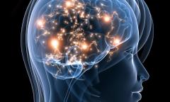
Neuroimaging
Neuroimaging enables us to look deep inside the brain to learn more about the impact of seizures on its structure and function.
Neuropathology
The Epilepsy Society Brain and Tissue Bank is the first of its kind in the UK. It is dedicated to the study of epilepsy through brain and other tissue samples.